The content on this page is intended to healthcare professionals and equivalents.
Produces consistent image quality across a wide range of patients.
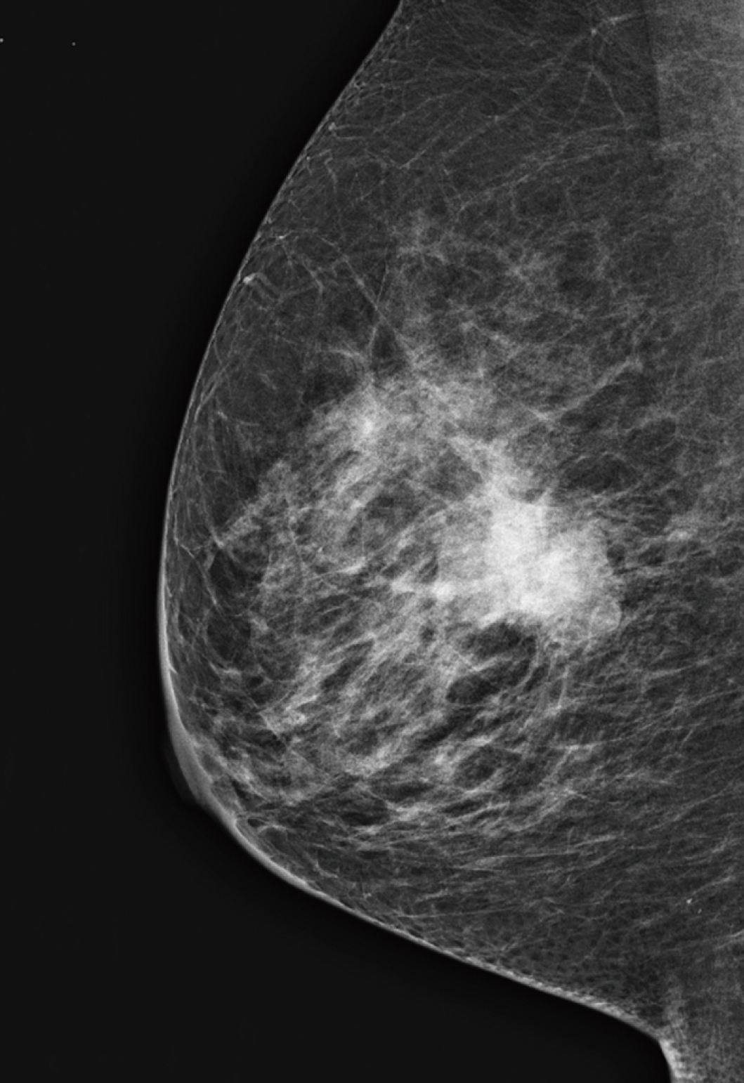
ASPIRE Cristalle incorporates adaptive image processing algorithms that automatically adjust to patient breast thickness and composition.
The detector is the heart of any digital mammography system. Simply put, its job is to detect x-rays, convert them into electrons, and collect the resulting electrical charges. The more efficiently it collects charges, the stronger the image signal, the less noisy the image, and the lower the dose needed.
In conventional detector design, the pixels that detect x-rays are square, with wide gaps between them, losing some of the converted x-ray information.
In the Aspire Cristalle, hexagonal pixels are arranged with smaller gaps between pixels, resulting in less signal loss, stronger electrical fields, and higher sensitivity. When compared to square pixels, HCP delivers:
- 20% increase in detector sensitivity
- Improved information capture
- Lower patient dose
- 50-micron display
- Fast acquisition time - only 15 seconds
- Hexagonal pixels distribute the electrical field more efficiently for a stronger, more homogenous signal
- Results in images with high DQE and MTF
- Ultra-sharp images, gentle dose
- Upgradeable to future technologies

Conventional square pixel

ASPIRE Cristalle hexagonal pixel
ISC-Image-based Spectrum Conversion is a contrast correction algorithm that produces molybdenum image quality even though the image was acquired with a tungsten target. ISC provides the dose savings of tungsten with the image quality of molybdenum.
Dynamic Visualization II (DYN II) provides consistent appropriate density of glandular and adipose tissue in each breast type, so the contrast of thick breast and dense breast is improved. Furthermore, it provides high contrast with no saturation in breast region, so the sites are possible to set high contrast parameter.

DYN II

MFP
Through the analysis of information obtained from low-dose, pre-shot images, Fujifilm's proprietary Intelligent AEC (IAEC) calculates the mammary gland density (breast type) and presence of implants when defining the x-ray energy and level of dose required.
IAEC optimizes exposure parameters. IAEC can also greatly speed workflow for exams of patients with Implanted breasts by automatically calculating the optimal exposure.
Automatically selects the appropriate mammary gland area from pre-shot images

Requires manual adjustment of the setting based on the assured location of mammary gland

Automatically selects the appropriate sensor from the pre-shot images
- * Some items are optional, please contact your subsidiary for the detail.


