The content on this page is intended to healthcare professionals and equivalents.
The advanced imaging technology for visualization of low velocity blood flow below the previous detection threshold. The unique algorithm displays fine blood flow with greater resolution and sensitivity.
Since its release in 2003, Real-time Virtual Sonography (RVS) has continued to evolve to meet clinical needs. Significant further developments have been introduced with the ARIETTA 850SE.

RTE assesses tissue strain in real time and displays the measured differences in tissue stiffness as a color map. Its application has been validated in a wide variety of clinical fields: for the breast, thyroid gland, and urinary structures.
Shear waves are generated using a ‘push pulse’ to excite the tissues. SWM provides an assessment of tissue stiffness by calculating Vs, the propagation velocity of the shear waves. SWM provides an additional reliability indicator, VsN, as an objective evaluation of the Vs measurement. SWE color-codes tissue sttifness based on the propagation velocity of shear waves. SWE can be used to evaluate liver visually and non-invasively.


By integrating the two non-invasive methods for evaluation of liver tissue stiffness, namely RTE and SWM, it is possible to assess the chronological progression of liver inflammation and fibrosis with greater accuracy. A combined simultaneous estimation of the degree of steatosis (ATT index) makes Combi-Elasto a comprehensive tool for the differential diagnosis of liver disease.
Three- and four-dimensional imaging can play a role as a prenatal communication tool connecting parents with their fetus. Auto Clipper automatically defines the optimal cut plane removing placental or other unwanted tissue signals in front of the fetus, offering a clear surface-rendered fetal image.
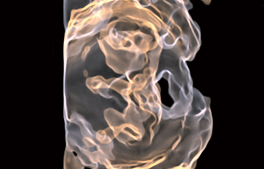
The 4Dshading technology gives a more realistic appearance to the rendered surface of the fetus in the 3D display. 4Dshading Flow is its Doppler blood flow mode optimized to offer a better understanding of complex vascular flows. 4Dtranslucence enables evaluation of fetal structures providing a display of the fetal body surface and internal organ boundaries with a translucency.


Automatic tracking of fetal heart movement from the B mode image follows the displacement of the heart wall in the apical direction for measurement of %Fractional Shortening (%FS). Measurement accuracy is unaffected by a change in the fetal position or by the mother’s breathing.
Enables observation of Doppler waveforms from two different locations during the same heart cycle. Simple measurements from two different waveforms can also be useful in the diagnosis of fetal arrhythmia.


