FUJIFILM

Verification of Healthcare Professionals

Thank you for visiting Fujifilm website.

The content on this page is intended for healthcare professionals or equivalent.
Please confirm that you are a healthcare professional.

You are accessing from the United States. To browse Fujifilm USA website, please click the following link.

Fujifilm USA Website

Clinical Applications

Abdomen Analysis - Features

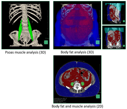
Calculates the area of the CT value corresponding to visceral fat and subcutaneous fat in 2D or 3D

The content on this page is intended to healthcare professionals and equivalents.

Body Fat Measurement

There are two types of obesity - visceral fat obesity and subcutaneous fat obesity and both can be determined with abdominal CT images. Visceral fat obesity leads to high risk of diseases such as cardiovascular diseases and lifestyle-related diseases hence early detection through medical examination is increasingly important.

Visual Explanation

Abdomen Analysis displays the areas corresponding to visceral fat and subcutaneous fat in different colors. Single-slice analysis as well as 3D analysis using multiple slices can be done. By displaying analysis results in 3D or graphically, details can be shown visually for easy understanding.

Simultaneous Analysis of Visceral Fat and Greater Psoas Muscle

In addition to visceral fat and subcutaneous fat, the area of the CT number corresponding to the greater psoas muscle and intramuscular fat can be calculated. 2D analysis by slices (slice processing) and 3D analysis using multiple slices (volume processing) are also available.

Volume Processing

The volume processing includes 3 types of display method for analysis results; “3D display”, “Graphical display” and “List display”. Display types can be switched with the tabs easily.

User-friendly Report Functions

In slice processing, an examination report to compare present and previous results and a mixed examination report on visceral fat analysis and greater psoas muscle analysis can be done.

In volume processing, report can be generated by setting image layout and selecting necessary analysis results. A list of analysis results can be also exported in the text format (CSV).

Analysis Results

Body fat analysis

  • Subcutaneous fat area [cm²] or volume [cm³]
  • Visceral fat area [cm²] or volume [cm³]
  • Abdominal circumference [cm]

Muscle analysis

  • Left greater psoas muscle area [cm²] or volume [cm³]
  • Right greater psoas muscle area [cm²] or volume [cm³]
  • Left erector muscle of spine [cm²] or volume [cm³]
  • Right erector muscle of spine [cm²] or volume [cm³]

Operating environment

  • Applicable system
    Whole-body X-ray CT system SCENARIA and Hyper Q-Net S
  • Report printing
    Internet Explorer (IE)
    Refer to the product specification for the IE version.
    Unable to print on the CT console.

* Please note that not all applications are available on all products listed. Please contact our sales representative to understand which products suit your needs.