The content on this page is intended to healthcare professionals and equivalents.
Detective Flow Imaging (DFI)

The advanced imaging technolgy for highly dynamic visualisation of low velocity blood flow below the previous detection threshold with high frame rate. The unique algorithm displays a clear and accurate information on blood perfusion with greater resolution and sensitivity.
Real-time Virtual Sonography (RVS)

Since its release in 2003, Real-time Virtual Sonography (RVS) has continued to evolve to meet clinical needs in fusion imaging and interventional procedure and open the way for Image-guided techniques and minimally invasive procedures. Significant further developments have been introduced with the ARIETTA 850.
Real-time Virtual Sonography (RVS)
3D Sim-Navigator

Provides simulation of single or multiple needle paths during navigation to a target with Real-time Virtual Sonography (RVS). The positional relationship between the marked target and needle paths can be assessed in real time using the 3D body mark, reconstructed from the virtual CT volume data, with additional C-plane display orthogonal to the needle path.
E-field Simulator

A color map superimposed on the CT image simulates the distribution of the electric field (E-field) from the given location of multiple electrodes during RFA treatment. The simulation can be made with different positions of the multiple electrodes to determine the optimal arrangement. This flexibility in planning the needle path can bring significant improvement to the treatment technique.
Elastography
Real-time Tissue Elastography(RTE)

Courtesy of: Norihiro Kokudo, M.D.,National Center for Global Health and Medicine
To understand tissue elasticity and differentiate between benign and malignant lesions RTE assesses tissue strain in real time and displays the measured differences in tissue stiffness as a color map. Its application has been validated in a wide variety of clinical fields: for breast, thyroid gland and urinary structures.
Shear Wave Measurement(SWM)/Shear Wave Elastography(SWE)
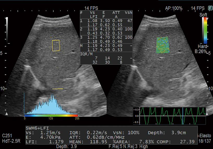
Avoid complex procedures like biopsy or MRI with our easy-to-use shear wave ultrasound features. Shear waves are generated using a ‘push pulse‘ to excite the tissues. SWM provides an assessment of tissue stiffness by calculating Vs, the propagation velocity of the shear waves. Thank to our unique reliability index, liver stiffness can be assessed with highly accurate quantitative information and reproducibility. SWE color-codes tissue stiffness based on the propagation velocity of shear waves. Both can be used to evaluate liver visually and non-invasively and enabling you to accurately stage the level of fibrosis.Furthermore, ATT measurement, to score the level of fatty infiltration, using the attenuation coefficient, is combined with Shear Wave Measurement for a complete multiparametric approach to chronic liver disease management.


Combi-Elasto

By integrating the two non-invasive methods for evaluation of liver tissue stiffness, namely RTE and SWM, it is possible to assess the chronological progression of liver inflammation and fibrosis with greater accuracy. A combined simultaneous estimation of the degree of steatosis (ATT index) makes Combi-Elasto a comprehensive tool for the differential diagnosis of liver disease.
Fetal 3D/4D

Three- and four-dimensional imaging can play a role as a prenatal communication tool connecting parents with their fetus. Auto Clipper automatically defines the optimal cut plane removing placental or other unwanted tissue signals in front of the fetus, offering a clear surface-rendered fetal image.
4Dshading Flow/4Dtranslucence
The 4Dshading technology gives a more realistic appearance to the rendered surface of the fetus in the 3D display. 4Dshading Flow is its Doppler blood flow mode optimized to offer a better understanding of complex vascular flows. 4Dtranslucence enables evaluation of fetal structures providing a display of the fetal body surface and internal organ boundaries with a translucency for an accurate assessment of fetal well-being from the very beginning.


Fetal Heart Examination
AutoFHR+

The fetal heart rate can be automatically calculated using a tracking ROI
placed over the fetal heart on the B mode image in real time. This offers a safer and more objective measurement compared to conventional Doppler or M-mode methods. Furthermore, as this function is also available on a transvaginal transducer, assessment can be made from early gestation onwards.
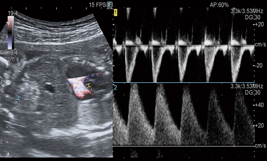
Dual Gate Doppler

Enables observation of Doppler waveforms from two different locations during the same heart cycle. Simple measurements from two different waveforms can also be useful in the diagnosis of fetal arrhythmia.


