This webpage content is intended for Healthcare Professionals only, not for general public.
Advanced Imaging-processing Technology for Versatile Diagnosis and Interventional Guidance
In the radiology field, high-level accuracy and reliability are necessary to ensure early detection, precise diagnosis, and appropriate treatment. Fujifilm has continued to develop advanced clinical technologies to facilitate fast and accurate examinations for you and your patients’ convenience.
Unique Flow Imaging

eFlow
High definition blood flow imaging mode with drastic improvement in spatial and temporal resolution. With higher sensitivity and less patient variability, it is possible to gain detailed observation and differentiation of fine blood vessels.

Detective Flow Imaging
Attain super visualisation of minute peripheral vessels and detect extremely low flow state with minimal background artefact.
Advanced Elastography Analysis
Tissue stiffness, or its elasticity, has long been known as a biomarker of tissue pathology. Ultrasound elastography was developed as a non-invasive approach to visualize tissue elasticity. By applying pressure to the human body through the transducer, the stiffness of a lesion can be evaluated to provide diagnostic information.

Real-time Tissue Elastography (RTE)
RTE assesses tissue strain in real time and displays the measured differences in tissue stiffness as a colour map. This gives greater visibility compared to B-mode image. Using the abdominal convex transducer, RTE can also provide an estimation of fibrosis staging in patients with hepatitis C (LF Index). This reduces the need for invasive procedures like biopsies, benefiting patients.
Its application has been validated in a wide variety of clinical fields: for the breast, pancreatic, thyroid glands and urinary structures.

Auto Frame Selection
To help maximize efficiency and reduce operator dependency, our systems automatically selects the optimum frames for measurement.

Assist Strain Ratio (ASR)
Fat Lesion Ratio (FLR), provides a quantitative method for the evaluation of regions of interest in the strain image. It compares the ratio between 2 ROIs; one on an area of fat, and another on the lesion being assessed.
Simply by clicking on the tumour, ASR is able to automatically set both ROIs necessary for FLR measurement, thereby improving the reproducibility and objectivity while shortening measurement time.

Shear Wave Measurement
Shear waves are generated using a 'push pulse' to excite the tissues. SWM provides an assessment of tissue stiffness by calculating Vs, the propagation velocity of the shear waves. Fujifilm's SWM provides an additional reliability indicator, VsN, as an objective evaluation of the Vs measurement.

Attenuation Index
Ultrasound attenuation gets higher as fat tissue increases. As such, the ATT value (attenuation measurement function) helps to assess hepatic fatty infiltration and it is not affected by liver inflammation and fibrosis.

Shear Wave Elastography
Based on the shear waves induced by ultrasonic beams, SWE can provide a quantitative estimation of tissue stiffness, and also display it as a colour-coded map, superimposed on the B-mode image in real-time. Selection of ROI is made easy with up to 5 flexible circle or ellipse measurements.

Combi-Elasto
The combined use of RTE and SWM offers a new approach to non-invasive assessment of liver fibrosis. It supports both conventional and quantitative indexes corresponding to fibrosis F value, inflammation A value, and fat amount ATT, to serve as a comprehensive tool to detail the chronological progression of liver diseases.
Guidance for Interventional Radiology

Real-time Virtual Sonography (RVS)
RVS merges a real-time ultrasound image with a previously acquired CT, MR, PET or ultrasound image. It uses the strengths of each imaging modality to give a direct comparison of lesions, useful from diagnosis to treatment to evaluation of the treatment program.

Micro Convex Transducer with Built-in Magnetic Sensor
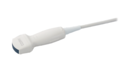
With our single crystal micro convex transducer, C23RV, there is no need for setting up an external magnetic sensor to kick start your RVS navigation. Simply plug in and you're ready to scan.

3D Sim-Navigator
Provides simulation of single or multiple needle paths during navigation to a target with Real-time Virtual Sonography (RVS).
The positional relationship between the marked target and needle paths can be assessed in real time using the 3D body mark, reconstructed from the virtual CT volume data, with additional C-plane display orthogonal to the needle path.

E-field Simulator
From the given location of multiple electrodes during RFA treatment, we are able to simulate a distribution of e-field as a colour map superimposed on reconstructed CT/MRI volume data. The simulation can be made with different positioning of multiple electrodes to determine the optimal arrangement. This flexibility in planning the needle path can bring significant improvement to the treatment technique.
Innovative Transducer
Matrix Linear Probe
The world’s first CMUT (Capacitive Micro-machined Ultrasound Transducer) probe in medical imaging with a super wide bandwidth (2-22MHz), it is a breakthrough innovation using the next generation silicon wafer technology. There is no matching layer required as silicon has an acoustic impedance close to human body. The ideal one probe solution that provides enhanced image resolution and high sensitivity for a wide range of ultrasound applications.


![[photo] DeepInsight - Redefining the way we see](https://asset.fujifilm.com/www/my/files/styles/1120x200/public/2024-09/e2951cfb180d4db419cf331f2bebf0e9/bnr_deepinsight_pc.jpg?itok=DF-Uqd16)