


The content on this page is intended to healthcare professionals and equivalents.
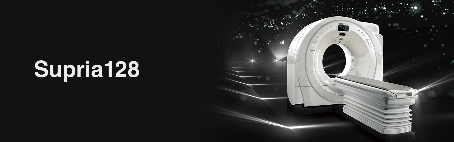
Supria 128

Due to the advance of medical care, CT device needs to be optimized for patient care and accelerate clinical decision-making more than before. On the other hand, the economical benefit is also demanded on medical finances due to rapidly growing aging society.
Supria128 has evolved in order to respond to the paradigm shift in the medical field with employing technologies such as whole-body submillimeter high-speed imaging and dose reduction technologies, which have become the "Next Standard" for challenging various clinical demands.
“Supria128” is a solution that responds to healthcare needs in the medical field.
“Supria128” Intends to Prove Lower Dose, High Quality Image
Iterative processing for routine examinations
Iterative Processing, used for dose reduction, requires plenty of calculation, making it difficult to apply it in routine examinations. In Supria 128, the image processing unit has been renewed and the processing speed improved, facilitating the use of Iterative Processing (Intelli IP) in routine examinations.
Optimal settings for each facility
Noise reduction strength can be selected from 7 levels. We provide high-quality images by reducing image noise and artifacts with an appropriate exposure dose according to the facility's operation policy.
Low tube voltage scanning
In general, low tube voltage scanning can be expected to increase CT values and improve low-contrast resolution with iodine contrast agents.The noise increased by low tube voltage imaging can be reduced by Intelli IP, which also reduces the burden on the patient.

“Supria128” Intends to Prove Lower Dose, High Quality Image
Equipped with a 0.625mm x 64ch = 40mm high resolution wide detector, submillimeter imaging is possible in a short scanning time at any region on the whole body.
Minimum slice width 0.625mm
The X-ray detector is separated by a partition. The X-ray utilization efficiency decreases by the thickness of this partition. There is a trade-off between X-ray utilization efficiency and spatial resolution, and a detector with a minimum slice width of 0.625mm is equipped.
Clinically effective
Since it is possible to take a wide range of scans, it is effective for various examinations such as head, lowered arms, and abdominal multi-phase scanning.

"Supria128" Intends to Prove Comfortable Patient Care
In-Room Operation
Scanning can be started / stopped on the gantry while monitoring the situation changes at a position close to the patient. This improves the workflow between the operation and scanning rooms, and consider the safety for smooth examinations.

High throughput, high image quality
High performance, such as high-speed rotation, submillimeter slice imaging, powerful X-ray generator and state-of-the-art image reconstruction algorithms, realizes high resolution and high throughput examinations.

High pitch scanning not limited to FOV
Our original 3D image reconstruction algorithm CORE method allows scanning with whole specification range of the slices thickness and FOV.
- * Depends on the specifications of the device
"Supria128" Intends to Prove High Functionality
HiMAR reduces metal artifacts

HiMAR (High Quality Metal Artifact Reduction) estimates and correctes artifacts based on metal data.
Helpful function for reducing the burden on the patient
Equipped with a motion artifact correction, body movement can be compensated even after scanning. Even if the patient is out of the effective field of view, such as a patient with a kyphosis, images can be reconstructed without re-scanning in case it is within the maximum effective field of view.
Motion artifact correction

Full FOV data acquisition

ECG Prospective scanning in synchronization with electrocardiogram
ECG Prospective scanning is a function that scans and achieves image in synchronization with electrocardiographic information. Images achieved by ECG Prospective scanning can be used for calcium scoring analysis*1.
- *1 A 3D workstation equipped with a calcium scoring analysis is required.
Capable of imaging in various patient's positions
With a large bore of 750mm and a maximum effective field of view that is patient friendly, it is possible to scan with various patient's positions.

Intuitive operability with Quick Entry
The scan button is located on the intercom box, just above the keyboard, with simply arranged operation buttons, large text, and an easy-to-understand display, which support examinations efficiently.

- * Fujifilm makes no representation that products on this website are commercially available in all countries.
- * Approved uses of products vary by country and region.
- * Specifications and appearance of products are subject to change without notice.
- * Please contact us for details


