


The content on this page is intended to healthcare professionals and equivalents.
LESS IS MORE
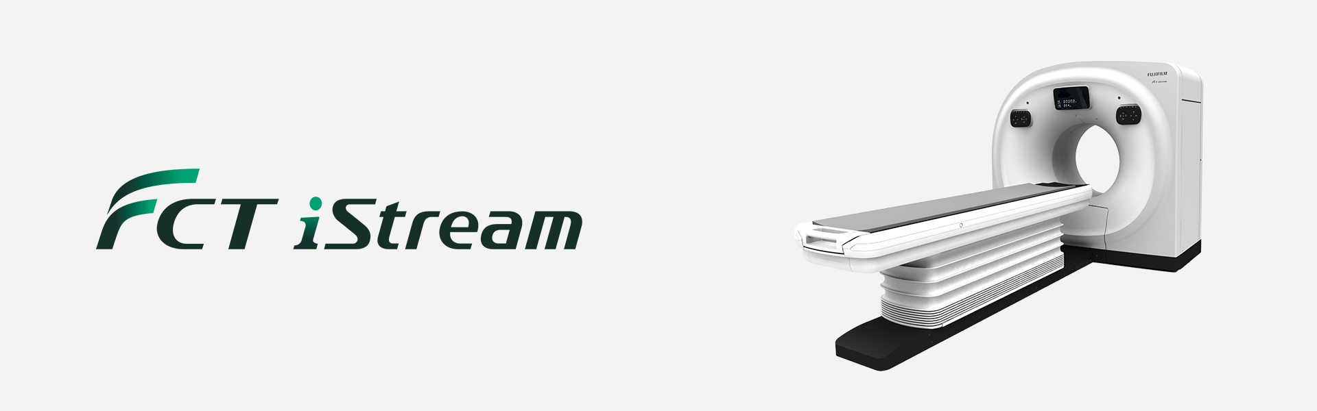
Technological innovations now deliver high-resolution, low dose images at high-speed, driving demand for X-ray CT systems in applications from prevention and diagnosis to treatment.
FCT iStream supports these CT applications with AI enhanced image processing and workflow to accommodate and address today’s increasing patient volume and dose concerns.

SynergyDrive is a generic term for technology related to workflow improvement. It includes functions developed by utilizing Machine Learning, an AI technology. The performance and accuracy of the system do not automatically change after implementation.
REiLI, FUJIFILM’s medical AI technology brand, enables support for physicians in the diagnosis and streamlining of the workflow for diagnostic imaging by combining the image processing technology we have cultivated with the most advanced AI technology to realize improved medical care.
- *1 Conventional CT system is our 64ch CT system without SynergyDrive.
SynergyDrive
EASE OF USE AND EFFICIENCY
01. Patient registration
Quick Entry Intuitive GUI design
Intuitive and easier operation with customizable GUI. Quick Entry mode enables simple operation for all users with fewer buttons and larger icons.
- CLICK
- CLICK
- PRESS "START" button

02. Positioning
One button setting
The table can be set to the pre-selected scan start position with one button on the head and body. Table height and table stop position can be customized according to the operation.

03. Scanogram

AutoPose & iTilt
FCT iStream automatically*2 sets the scanning range using the scanogram image by AutoPose function. Since the margins can be set in advance, the range can be customized according to the preference of the facility. iTilt automatically creates and enables observation of tilted images during scanning.

When set to OM Line

When set to SM Line

When set to RB Line

chest
Red: automatically set position, Blue: automatically set position + margin setting position
04. Scan
60ips high-speed image reconstruction
Image reconstruction is performed at a maximum speed of 60 ips in parallel with scanning (data acquisition). Images can be checked during scanning, enhancing workflow.

05. Processing/Transfer
Image transmission to Synapse 3D in parallel
The reconstructed image is displayed on the console and also saved in SYNAPSE 3D in parallel. Eliminates the need to send DICOM images from the console and improves the operability of examinations.

- *2 The automatic selection must be checked and the operator may adjust, if necessary.
LOW DOSE,
HIGH QUALITY IMAGE
Intelli IPV
Our iterative reconstruction method, Intelli IPV, provides images that maintain their natural texture even at high noise reduction rates and their excellent visibility even at low doses, and does not require a dedicated operating room or additional hardware.

While adjusting the texture at a uniform ratio from high frequency to low frequency, the physical properties that affect visibility are made as close as FBP.

- * IPV stands for Iterative Progressive reconstruction with Visual modeling.
- Intelli IPV was developed using Machine Learning, an AI technology. The performance and accuracy of the system do not automatically change after implementation.
IntelliEC Plus
IntelliEC Plus is the auto exposure control function considered with iterative reconstruction parameters. It supports accurate low-dose scanning with simple operations.

IntelliODM
IntelliODM organ-based tube current modulation reduces dose from the anterior side where locate radiation sensitive anatomy such as lenses as much as 30% without any image quality compromise.


HiMAR Plus
HiMAR Plus reduces metal artifacts using an iterative processing to estimate the artifact. HiMAR Plus strength can be varied according to the application.
![Clinical images [HiMAR Plus]](https://asset.fujifilm.com/www/ae/files/2024-08/037d1f7404e31b7e9d191cd7c0a1e919/pic_fct-istream_ov_13.png)

- *3 It is obtained in the abdominal region.
- *4 Compared to FBP. It was measured using Intelli IPV intensity level Strong5 and tested to a water phantom. Depending on the clinical task, patient size, anatomic location, and clinical examination, the effect obtained may be smaller.
- *5 Compared to FBP. It was measured at 0.625 mm slice thickness using Intelli IPV intensity level Strong5 and tested to MITA CT IQ phantom CCT189, Phantom Laboratory using the model observer method results. Depending on the clinical task, patient size, anatomic location, and clinical examination, the effect obtained may be smaller.
EASIER TO USE
WIDER USAGE
IntelliEC Cardiac
IntelliEC Cardiac reduces dose using electrocardiogram information. Dose modulation can be applied to minimize tube current during non-target phases, including resting phases of mid-diastole and end-systole.


Reconstructed cardiac phase 75%
CardioHarmony
CardioHarmony enhances cardiac CT workflow by automatically*6 detecting and reconstructing cardiac phases with minimal motion.


- *6 The automatic selection must be checked and the operator may adjust, if necessary.
EASY TO INSTALL
EASY TO MANAGE
3-Unit Design Compact Space
The entire system comprises just three components; gantry, patient table, and operator console. No additional units such as the system transformer are required, so the space in the CT room can be maximized for clinical use.

Eco mode
Eco mode has two functions (On-time Standby and Off-time mode). The On-time Standby function controls the units mounted in the gantry, thereby reducing power consumption by up to 41% compared to Eco mode OFF. The Off-time mode reduces the electricity consumption of the detector while maintaining the operating readiness of the detector when the system is power-off. With Off-time mode, the power consumption during standby is reduced by up to 62% compared to when not in use.

Dose Management for various purposes
Simple Dose Report
Dose information can be transferred to the PACS as a secondary image. It enables the confirmation of the dose information on the PACS Viewer same as DICOM images.

Report example

DICOM SR
Dose information can be sent to PACS, etc. as a DICOM structured report (DICOM SR) along with the DICOM standard.
- * Fujifilm makes no representation that products on this website are commercially available in all countries.
- * Approved uses of products vary by country and region.
- * Specifications and appearance of products are subject to change without notice.


